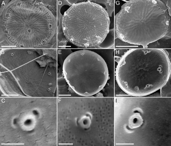
Figure 3.
Scanning electron micrographs of three ecologically diverse representatives of Thalassiosira pseudonana. (A-C) Marine culture strain CCMP1335 whose genome was sequenced; (D-F) freshwater specimens from the original type collection for the species from the River Wümme, Germany; (G-I) freshwater culture strain ETC1 from Lake Erie, Michigan, used in molecular phylogenetic analyses (Figure 1). The first and second rows show the cell exterior and interior, respectively (scale bar = 1 μm), and the third row shows the interior ultrastructure of the strutted process (scale bar = 200 nm).

