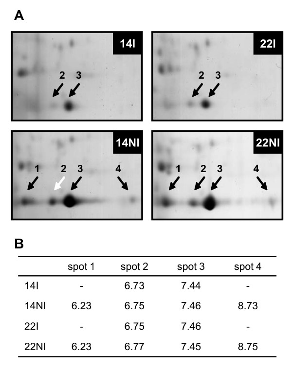
Figure 6.
Differential accumulation of RBCS subunits in leaves of C. canephora subjected to different water regimes. A: Proteins were extracted from clones 14 and 22 grown with (I) or without (NI) irrigation (14I, 14NI, 22I and 22NI) and analysed by two-dimensional gel electrophoresis (2-DE). Only parts of the 2-DE gels containing RBCS proteins are shown. Black arrows indicate RBCS spots. The RBCS1 protein analysed by MS/MS is shown by a white arrow. B: Isoelectric points (pI) of RBCS proteins identified by 2-DE gel electrophoresis. The pIs of were determined from calibrated 2-DE gels using ImageMaster Platinum 6.0 Software. The absence (-) of a pI value indicates that the isoform was not present in the gel.

