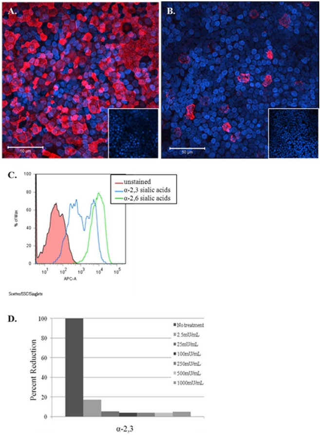
Figure 1. Fully differentiated NHBE cells express both α2,6 and α2,3 sias.
NHBE cells were stained for α2,6 (A) or α2,3 (B) sias shown in red. Cells pre-treated with neuraminidase abolished sias residue staining (image inserts). (C) NHBE cells were trypsinized, fixed with 2% formaldehyde, and analyzed by flow cytometry to determine relative percentage of cells staining positive for α2,3 (blue), or α2,6 sia moieties (green). The x-axis shows the mean fluorescence intensity and the y-axis shows the percent positive staining cells. Results shown are representative of four independent experiments. (D) NHBE cells were trypsinized, treated with the indicated concentrations of neuraminidase, and analyzed as in (C) to determine the percentage of cells staining positive for detectable α2,3 sialic acid residues. Results shown are representative of two independent experiments.

