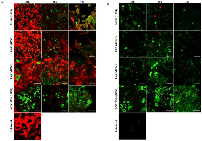
Figure 4. AIVs infect NHBE cells independent of α2,3 sias expression.
NHBE cells were infected with MI/06, PA/93, TX/02 or NY/04 at MOI = 0.5. At the times indicated post-infection, cells were fixed with 3.7% formaldehyde in PBS for 30 minutes. Cells were stained for α2,6 (A) or α2,3 (B) sialic acids (red) and influenza NP (green). Results shown are representative of three independent experiments.

