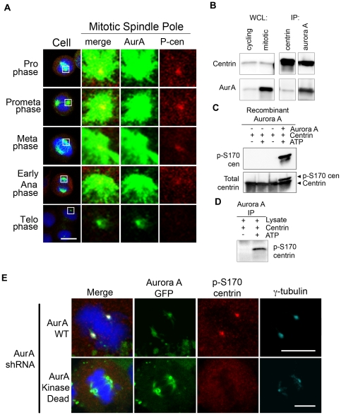
Figure 1. Aurora A localizes with and phosphorylates centrin in vitro and in cells.
(A) Immunofluorescence confocal microscopy of HeLa cells demonstrates that Aurora A (green) and phospho-S170 centrin (red) both localize at mitotic spindle poles from prophase through metaphase. Phospho-centrin is greatly reduced in anaphase and telophase and Aurora A is greatly reduced by telophase. DNA was counterstained with DAPI (blue). Scale bar = 10 microns. (B) Western blots of whole cell lysates (WCL) from cycling and nocodazole-arrested mitotic cells show that centrin and Aurora A are present in asynchronous and synchronous cultures, with greater amounts of Aurora A in mitotic cells. Reciprocal immunoprecipitations (IPs) of mitotic cell lysates demonstrate an interaction between Aurora A and centrin during mitosis. (C) In in vitro kinase assays with recombinant centrin incubated with recombinant Aurora A, centrin was phosphorylated only in the presence of ATP. Under conditions containing ATP, a shift in centrin is detected when Western blotted with a total centrin antibody (lower panel) and phospho-centrin is detected by the phospho-S170 centrin antibody (upper panel). Phospho-centrin is not detected under conditions lacking Aurora A and/or ATP. (D) In vitro kinase assays using endogenous Aurora A immunoprecipitated from nocodazole-arrested HeLa cells generated phospho-centrin only in the presence of ATP as demonstrated by Western blotting with the phospho-S170 centrin antibody. (E) Kinase-active Aurora A is required for phosphorylation of centrin at mitotic spindle poles. Cells that lack endogenous Aurora A but express shRNA-resistant GFP-WT Aurora A clearly exhibit phospho-S170 centrin (red) staining; while those that lack endogenous Aurora A and express GFP-kinase dead Aurora A exhibit a nearly complete loss of phospho-S170 centrin (red) staining at the mitotic spindle poles. White arrows denote the focal staining of gamma-tubulin (turquoise) at poles in the cell expressing WT Aurora A or kinase dead Aurora A (green).

