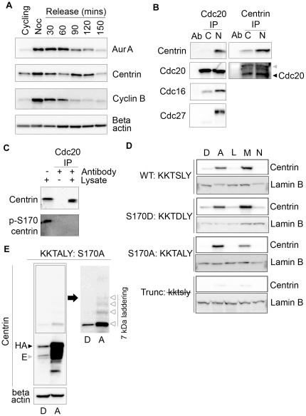
Figure 3. Centrin interacts with APC/C during mitosis.
(A) Levels of Aurora A, centrin, and cyclin B were compared in Western blots of lysates of HeLa cells harvested at the indicated time points after synchronization by double thymidine/nocodazole block and release. Cyclin B degradation is used to indicate the onset of anaphase, while the beta-actin blot serves as a loading control. Aurora A and centrin levels drop to basal levels by 150 minutes post-release, while cyclin B is at basal levels by 120 minutes post-release. (B) Equal volumes of immunoprecipitations of lysates from cycling (C) or nocodazole arrested (N) HeLa cells performed with Cdc20 and centrin and Western blotted with the indicated antibodies demonstrate that centrin is pulled down with cdc20 only in nocodazole arrested cells along with Cdc16 and Cdc27. Cdc20 is pulled down with centrin in nocodazole arrested cells and to a slightly lesser extent in cycling cells. The antibody heavy chain and cdc20 were indicated with ( ) and (◀), respectively. (C) In lysates from asynchronously growing HeLa cells only non-phosphorylated centrin immunoprecipitates with cdc20. Cdc20 does not pull down phosphorylated centrin even though p-S170 centrin is abundant, as seen in the lysate-only lane. (D) HeLa Tet-On cells expressing various centrin mutants treated overnight with DMSO (D), ALLnL (A), leupeptin (L), MG132 (M), and ammonium chloride (N) and Western blotted with antibodies directed against total centrin reveal that DMSO, leupeptin, and ammonium chloride do not prevent centrin degradation, whereas ALLnL and MG132, the two proteasome inhibitors, do prevent degradation of wildtype and mutant forms of centrin. Lamin B loading controls are shown for each mutant cell line. (E) Lysates from HeLa Tet-On cells expressing S170A centrin treated with DMSO (D) or ALLnL (A) for 16 hours were Western blotted for centrin and beta-actin show significant degradation products in the presence of ALLnL but not DMSO. Additionally, when the boxed area of the centrin blot is over-exposed, 7 kDa laddering indicative of ubiquitination is evident (Δ). Endogenous and HA-centrin are indicated with (
 ) and (▶), respectively.

