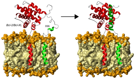
Figure 9. Tentative structural model of Harakiri's operating mode.
The survival protein (Bcl-2 or Bcl-xL) [13], [14] and Hrk are represented as red and green ribbons, respectively. The arrow connects the states before (Hrk's cytosolic domain is shown largely disordered with preformed secondary structure) and after (Hrk's cytosolic domain is forming a helix) the interaction between Hrk and the survival partner. The hydrophobic and polar parts of the bilayer are colored in light and dark yellow, respectively.

