
FIG. 14.
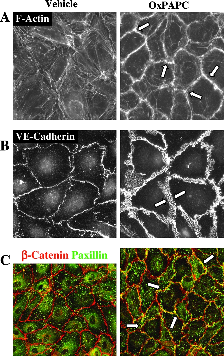
OxPAPC-induced endothelial remodeling. Enhancement of peripheral endothelial actin cytoskeleton (A, arrows), VE-cadherin positive adherens junctions (B, arrows), and peripheral colocalization of focal adhesions and adherens junctions (C, arrows) detected by double immunofluorescent staining for β-catenin (red) and paxillin (green) and confocal microscopy. ECs were stimulated with barrier-protective OxPAPC concentration (20 μg/ml). Adapted from (31, 33).