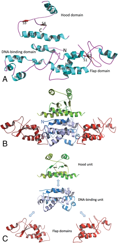
Fig. 2.
HetR structure. (A) The HetR subunit B fold. α-Helices (H) are in aqua; β-strands (S) are red, and loops are purple. N and C termini are labeled. (B) Crystal structure of the HetR dimer. A ribbon representation of the HetR structure. Secondary structure elements are indicated by H (helices) and S (β-strands). Subunits A and B are shown in lighter colors (light red, blue, and green) and darker colors (dark red, blue, and green), respectively. The DNA-binding unit is shown in blue, flap domains in red, and the hood in green. Protein N and C termini are labeled. (C) Domain organization of the HetR dimer.

