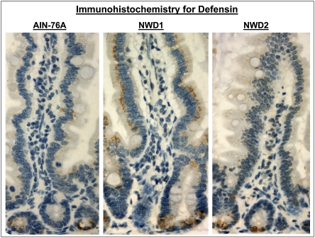
Fig. 4.
Immunohistochemical detection of the Paneth cell marker defensin in the intestinal mucosa of mice maintained on AIN76A, NWD1, or NWD2 for 1 y from weaning. Positive staining is brown and localized to Paneth cells in the crypt for mice fed AIN76A or NWD2, but also, it is present in the apical region of normal-appearing enterocytes in mice fed NWD1.

