Fig. 5.
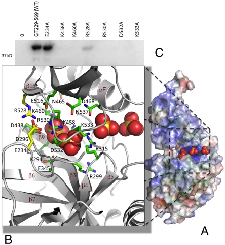
Active site cleft of hGTase. (A) Four sulfate ions [in Corey–Pauling–Koltun (CPK) model] are bound at the active site cleft of one of the hGTase molecules; the molecule is viewed at approximately 70° clockwise rotation about the y axis with respect to the view in Fig. 4. The amino acid residues surrounding the sulfate ions are highly conserved and, if mutated, impair or eliminate GTase activity in human (Table 1) and mCE (44). (C) hGTase containing E234A and R528A mutations (positions shown as yellow side-chains in B) retained full and 10% activity, respectively.

