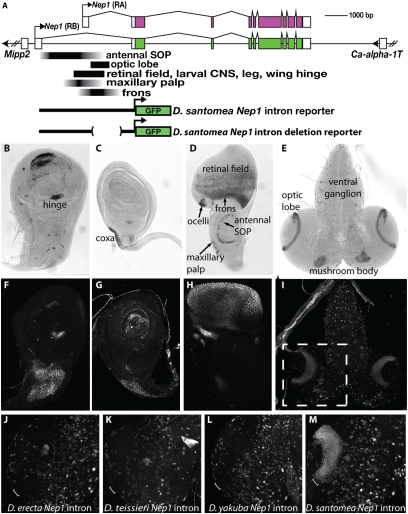
Fig. 2.
Mapping of Nep1 enhancers reveals that the novel optic lobe expression pattern evolved through changes in cis to Nep-1. (A) Schematic of the Nep1 locus showing the location of enhancers that were mapped in the first intron of D. santomea and reporter constructs used in the study. Nep-1 mRNA expression within third instar larval imaginal tissues of D. santomea was revealed by in situ hybridization to a wing disc (B), leg disc (C), eye-antennal disc (D), and larval brain (E). Expression of a D. santomea Nep1 intron-GFP reporter fusion construct in larval imaginal tissues recapitulates patterns of endogenous Nep1 expression in the wing hinge (F) and leg coxa (G) as well as a complex ensemble of patterns in the eye-antennal disc (maxillary palp, retinal field, ocelli, and frons) (H). Expression patterns in the larval brain, including cells of the ventral ganglion, mushroom bodies, and the D. santomea-specific expression in laminar neuroblasts of the optic lobes (E), are governed by the D. santomea Nep1 intron (I). Dashed box in I delimits the optic lobe region presented in J–M. (J–M) Reporter expression in the optic lobe driven by Nep1 intron constructs derived from multiple species. Optic lobes of D. melanogaster transgenic for Nep1 intron reporter constructs derived from D. erecta (J), D. teissieri (K), and D. yakuba (L) lack the activity in laminar neuroblasts that is driven by the D. santomea intron (compare white bracket in M with J, K, and L).

