Fig. 1.
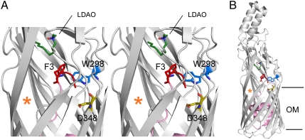
The N terminus of FadL is connected to the high-affinity binding site. (A) Stereo cartoon viewed from the side, showing the locations of F3, W298, and D348 within the wild-type EcFadL structure (Protein Data Bank ID 1T16). The N terminus (residues 1–3) is colored red, with the rest of the plug domain colored pink. The LCFA-mimicking LDAO detergent molecule bound in the high-affinity binding site is colored dark green (nitrogens blue, oxygens red). The lateral opening in the barrel wall is indicated with an orange asterisk. Residues 405–411 in strand S14 have been removed to show the LDAO detergent molecule more clearly. The extracellular side is at the top of the figure. (B) Overview showing the complete EcFadL structure for reference, with the hydrophobic core of the OM indicated as horizontal lines. This and the following figures were made with PyMOL (14).

