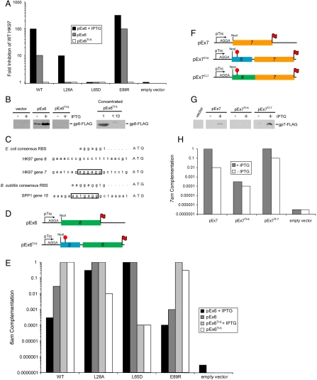
Fig. 2.
Analysis of the expression of HK97 connector proteins. (A) Inhibition of WT HK97 phage plating efficiency resulting from expression of gp6 from the indicated plasmids. (B) Plasmid-based expression levels of gp6. Lysates made from cells carrying the indicated plasmids were separated by SDS-PAGE and immunoblotted with an anti-FLAG antibody (Left). gp6 produced from pEx6TI-6 was concentrated by Ni-NTA chromatography [2,000-fold relative to samples analyzed (Left)] and dilutions of this preparation were analyzed by SDS-PAGE and immunoblotting (Right). (C) The nucleotide sequences upstream of HK97 genes 6 and 7, and SPP1 gene 15. Ribosome binding sites are boxed. Consensus ribosome binding sites for the hosts of these phages [E. coli (34) and B. subtilis (35)] are also shown. (D) Schematic representations of gp6 constructs used in this work. pEx6 contains gene 6 (green) under the translational control of the strong ribosome binding site encoded within the parent expression vector. pEx6TI-6 contains 150 bp upstream of gene 6, which includes a portion of gene 5 (blue) cloned out of frame with respect to the plasmid encoded translation start (red “stop” sign). Translation of gene 6 on this vector is mediated by its native translation initiation site. Both vectors express gp6 with a C-terminal FLAG epitope (red flag). (E) Complementation of an HK97 6am phage by WT and mutant forms of gp6 expressed from the indicated constructs. Values were normalized to the complementation mediated by WT gp6 expressed from pEx6TI-6. (F) Schematic representations of gp7 constructs used in this work. For pEx7, gene 7 (orange) is expressed under the translational control of the plasmid-based translation initiation site as in pEx6. pEx7TI-6 is constructed similarly to pEx6TI-6 such that translation of gene 7 is mediated by the native translation initiation site of gene 6. In pEx7TI-7, the translation of gene 7 is mediated by its native translation initiation site. (G) Expression level of gp7 in cells carrying pEx7, pEx7TI-6, and pEx7TI-7. These experiments were carried out as described in panel 2B. (H) Complementation of an HK97 7am phage by gp7 expressed from the indicated construct. Values were normalized to the complementation mediated by gp7 expressed from pEx7TI-7 in the presence of IPTG.

