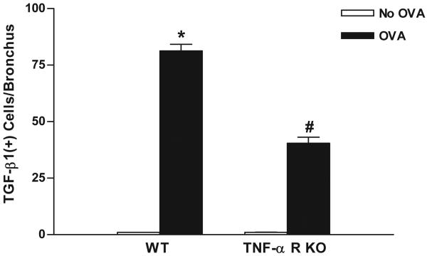
Figure 4. Levels of peribronchial TGF-β1+ cells in TNF-R deficient vs WT mice.
TNF-R deficient or WT mice were subjected to chronic OVA challenge. Non-OVA challenged mice served as a control. Lung sections were immunostained with an anti-TGF-β1 Ab and the number of peribronchial TGF-β1+ cells determined by image analysis. Chronic OVA challenge in WT mice induced a significant increase in the number of peribronchial TGF-β1+ cells (p<0.0001*)(WT no OVA vs WT OVA). The number of peribronchial TGF-β1+ cells were significantly reduced in chronic OVA challenged TNF-R deficient mice compared to WT mice challenged chronically with OVA (p<0.0001#)(n=16 mice/group).

