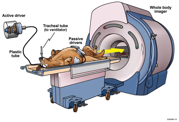
Figure 3. Magnetic Resonance Elastography in a Large Animal Model.
The MRE apparatus is illustrated including a mechanical system for application of shear waves through the abdomen. The animals were intubated and kept under general anesthesia. Two small separate drivers were strapped around the abdomen (as opposed to one large driver applied to the abdomen of humans) due to the V-shaped form of the canine torso. During data aquisition, acoustic pressure waves (60 Hz) were generated by an active pneumatic driver located outside of the magnetic field of the whole body imager and conveyed via a flexible tube to the two passive pneumatic drivers strapped around each side of the upper abdomen. MRE, Magnetic Resonance Elastography.

