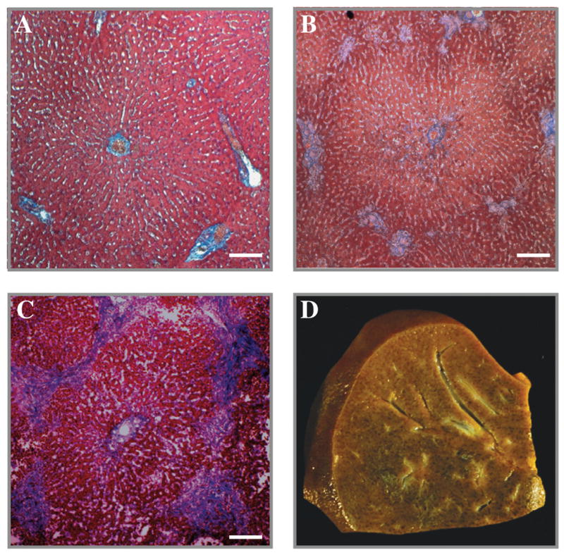
Figure 5. Liver Histology.

Low power magnification of canine liver stained with Masson’s trichrome at baseline (A), four weeks (B), and eight weeks (C). Note the mild patches of blue staining around the portal triads indicating an F1 fibrosis after four weeks (B), and progression to bridging fibrosis (F3) after eight weeks (C). A nutmeg appearance of the sliced liver characteristic of cholestatic liver disease was observed after eight weeks (D).
