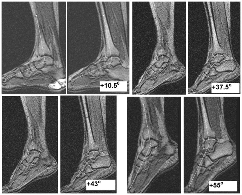
Figure 2.

Anatomical images of the ankle acquired in the body coil at four ankle positions with ankle angle as the average of the measurements in two slices at each position. Positive ankle angle values shown in the images are the deviations from the neutral position (00) in the plantarflexed direction. This imaging based assessment allowed accurate positioning and determination of the ankle angle.
