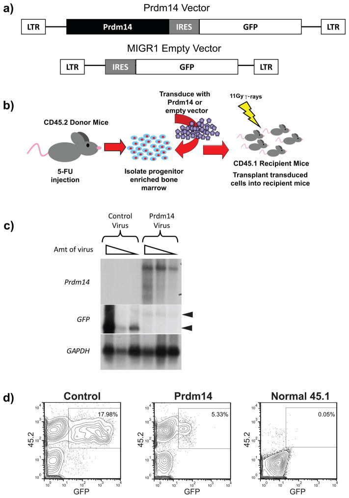
Figure 1. Overexpression of Prdm14 in vivo.
1a) Murine stem cell virus vectors were used to overexpress Prdm14 and serve as an empty vector control. Both vectors contain an IRES sequence and express GFP as a marker of transduced cells. 1b) An overview shows the experimental method used to express Prdm14 in vivo. Mice expressing the CD45.2 cell surface antigen are treated with 5-fluorouracil (5-FU) to induce cycling and enrichment of BM hematopoietic stem cells (HSCs). These CD45.2 donor cells are transduced with retroviral particles containing either Prdm14-MIGR1 or the EV-MIGR1 control and then injected intravenously into lethally irradiated recipient mice that express the CD45.1 allele. Engraftment of transduced cells allows for long-term overexpression of Prdm14. 1c) Northern blot analysis of transcripts produced from NIH3T3 cells transduced with Prdm14-MIGR1 or the control vector. A GFP probe detects both constructs and shows a higher level of expression in cells transduced with the control vector than with the Prdm14-MIGR1 vector. A second probe detects the Prdm14 transcript only. Both vectors produce transcripts of the expected sizes, shown as arrowheads. 1d) Four weeks after transplantation, fewer GFP+ cells are detected in PB of mice transduced with Prdm14-MIGR1 than with the control. A normal CD45.1 mouse has no GFP fluorescence.

