Fig. 1.

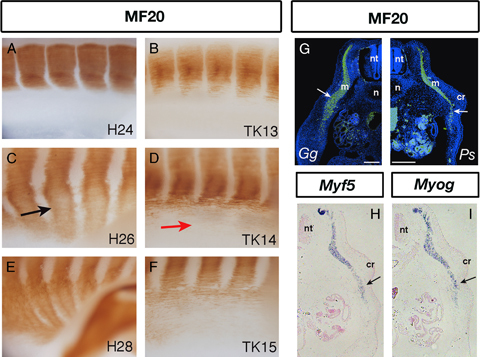
Comparison of muscle plates between Pelodiscus sinensis and chicken embryos. Whole-mount immunostaining of myosin heavy chain using the MF20 antibody in chicken and P. sinensis embryos. Hamburger–Hamilton (HH) stages 24 (A), 26 (C), and 28 (E) chicken embryos and Tokita–Kuratani (TK) stages 13 (B), 14 (D), and 15 (F) P. sinensis embryos were examined. The black arrow in (C) indicates muscle plate tissue extending into the lateral body wall region, which maintains its segmental organization. The red arrow in (D) shows MF20-positive myotomal fibers in the lateral body wall in a P. sinensis embryo. (G) Transverse sections immunostained with MF20. Note the massive and tightly packed muscle plate in the lateral body wall of an HH stage 26 chicken embryo (left), compared with the sparse myotomal cells in a TK stage 14 turtle embryo muscle plate (right). (H, I) Section in situ hybridization of TK stage 14 P. sinensis with a probe for Myf5 (H) and myogenin (I). Arrows in (G), (H), and (I) indicate the junction of the lateral body wall and the axial part of the embryonic body. Scale bar=100 μm for (G). cr, carapacial ridge; m, myotome; n, notochord; nt, neural tube.
