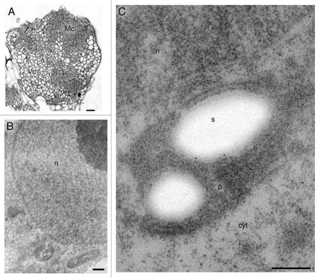
Figure 3.
Anti-ADC immunogold labeling in meristematic nodular cells after 28 days of culture. (A) Semi-thin section showing meristematic (Mc) and vacuolated cells (Vc) of nodules. (B) Magnification of meristematic cells. (C) Meristematic cell showing gold particles over nucleus, cytoplasm and plastids. Bars: A = 100 µm; B = 400 nm; C = 300 nm.

