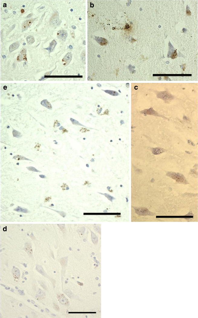
Fig. 2.
PIN1 granules in other disorders and in ND cases. The appearance of PIN1 granules is consistent in different groups and similar to those in a (a–d), except in FTLD cases where PIN1 granules show weaker immunoreactivity (c). There is preferential involvement of CA2 in PSP (a), FTLD (c) and PD (d). b PIN1 granules in CA4 in MND. e Involvement of CA1 in ND cases. All scale bars 20 µm

