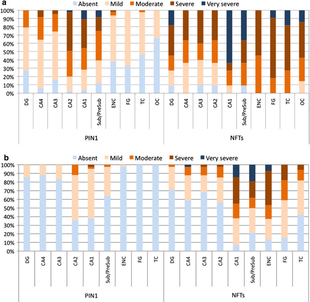
Fig. 5.
The distribution of PIN1 granules and NFTs in AD (a) and in PD/DLB (b). PIN1 granules occur more severely in CA1 and CA2 subfields and the subiculum in AD (a) but less so in cortical regions; a topographical pattern was also reflected in PD/DLB cases, though much less severely so (b). NFT distribution is independent of the distribution of PIN1 granules with NFT pathology occurring less severely in CA2 subfield and more frequently in cortical regions. Results are expressed as percentages of cases. DG dentate gyrus, Sub/Presub subiculum/presubiculum, ENC entorhinal cortex, FG fusiform gyrus, TC temporal cortex

