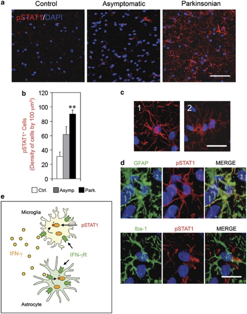
Figure 4.
Persistent phosphorylation of STAT1 (pSTAT1) in the SNpc of chronic Parkinsonian monkeys. (a–e) Panel (a) shows confocal images of pSTAT1 in a representative image of the SNpc of a control monkey, asymptomatic and Parkinsonian monkey. DAPI staining is combined to observe the cell nuclei (Blue). High levels of pSTAT1 staining (Red) can be seen in Parkinsonian monkeys. Scale bar: 100 μm. (b) Quantification of pSTAT1+ cells in the SNpc of control, asymptomatic and Parkinsonian monkeys. A strongly significant increase can be observed in Parkinsonian monkeys. **P<0.01 (one-way ANOVA and Tukey's test). (c) The pSTAT1 immunostaining in the SNpc identified astroglia-like cells (1) and microglia-like cells (2). Scale bar: 30 μm. (d) The pSTAT1 immunostaining (Red) was combined with GFAP or Iba-1 antibodies (Green) and both astrocytes (GFAP) and microglia (Iba-1) were seen to co-localize with pSTAT1. Nuclei were stained with DAPI (Blue). Scale bar: 20 μm. (e) Illustration of the IFN-γ signaling, characterized by the phosphorylation of STAT1, through the activation of IFN-γ receptor (IFN-γR) in a microglial cell and an astrocyte

