Figure 1.
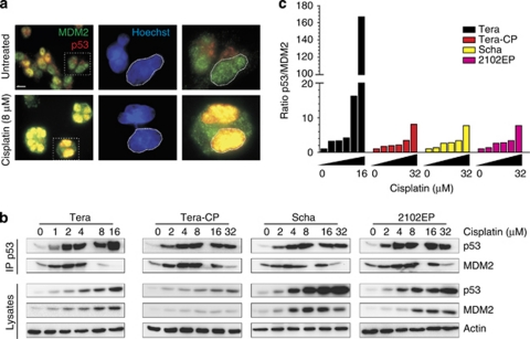
Earlier onset of cisplatin-induced loss of p53–MDM2 complex formation in cisplatin-sensitive TC cells. (a) Immunofluoresence showing that p53 becomes more nuclear localised, whereas MDM2 becomes nuclear and cytoplasmic localised after cisplatin treatment in the cisplatin-sensitive TC cell line Tera, representative example of three independent experiments. Selected area of the original image, as indicated, × 4 digitally magnified. Scale bar: 30 μM. (b and c) Note that we have used lower cisplatin concentrations for Tera compared with the other TC cell lines. TC cells were harvested 12 h after indicated cisplatin treatment. (b) Cell lysates were subjected to p53 IP. Immunoblotting was performed using anti-p53 and anti-MDM2 antibodies. In the cisplatin-resistant TC cell lines Tera-CP, 2102EP and Scha, p53 is maintained in a complex with MDM2 after cisplatin treatment, while the cisplatin-sensitive Tera cells show a loss of p53–MDM2 complex formation at low cisplatin concentrations. (c) Relative levels of p53 and MDM2 were calculated with imageJ 1.41 (National Institutes of Health, http://rsbweb.nih.gov/ij/index.html), normalised and divided p53/MDM2

