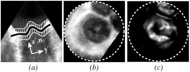
Fig. 5.
(a) Slice normal to the valve plane from 3DUS of a prolapsed mitral valve (same data as shown in Fig. 2) showing MVsurf (black) and adjacent regions (white striped) used to form Pint, (b) intensity projection Pint, and (c) thin tissue detector projection Pttd. Projection images are only defined within the dotted circles.

