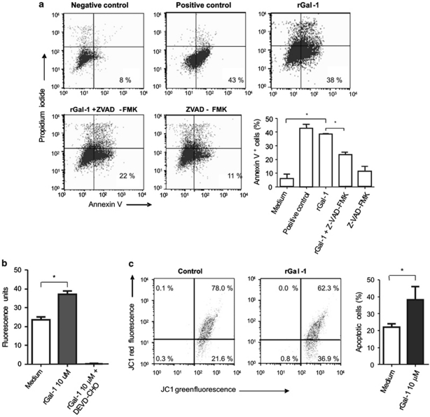
Figure 3.
Mechanisms underlying Gal-1-induced enterocyte apoptosis. (a) Flow cytometry analysis of annexin V/PI staining of enterocytes cultured for 16 h with different stimuli. Enterocytes were incubated with medium plus FBS (negative control), with medium without FBS (apoptosis positive control, with 10 μM rGal-1, with rGal-1 10 μM before incubation for 2 h with ZVAD-FMK (a pan-caspase inhibitor) or with medium before incubation for 2 h with ZVAD-FMK. Results are from one representative of four independent experiments. The accompanying graph shows average percentage of annexin V+/ PI− cells from all experiments. Statistically significant differences of P<0.05 are denoted as starred values (*). (b) Enzymatic activity of caspase-3 was determined with the caspase-3 specific fluorometric substrate Ac-DEVD-AFC. Enterocytes were incubated with 10 μM rGal-1 for 16 h and then harvested, lysed and analyzed. DEVD-CHO is a caspase-3-specific inhibitor. Data are results of four independent experiments (mean value±S.E.M.). *P<0.05. (c) Alteration of the mitochondrial membrane potential in enterocytes incubated for 16 h with medium alone (negative control, left dot plot) or with 10 μM rGal-1 (right dot plot). Data are results of four independent experiments. *P<0.05

