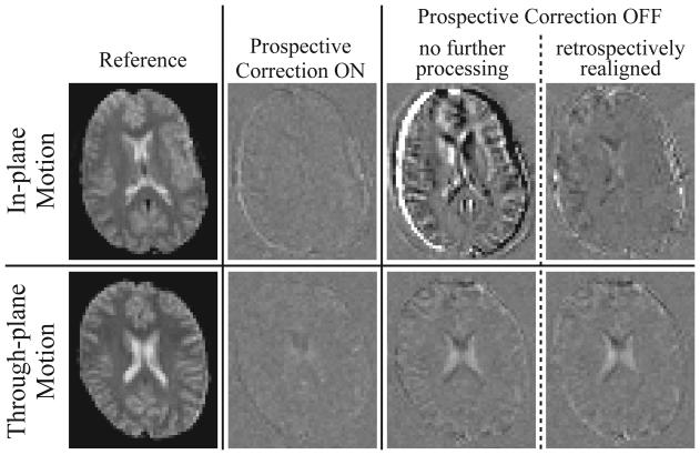
Fig. 2.
Difference images (Iafter - Ibefore) obtained from one volunteer performing a continuous in-plane motion (row 1), and a second volunteer performing a continuous through-plane motion (row 2). A zero difference image reflects perfect correction. A reference EPI image (column 1) is shown alongside sample difference images generated from time-series acquired with prospective correction ON (column 2) and OFF (columns 3,4); column 3 images remained completely unprocessed, while column 4 images were retrospectively realigned using SPM. Row 1 was chosen to demonstrate an in-plane motion case with a large rotational displacement θs and moderate intra-volume motion Δθs; the closest matching slices with prospective correction ON (row 1, column 2: Iafter acquired at θs = 3.52°, Δθs = 1.25°) and OFF (row 1, columns 3,4: Iafter at θs = 4.38°, Δθs = 1.07°) are chosen for comparison. Row 2 was chosen to demonstrate through-plane motion with a small rotational displacement θp but a large intra-volume motion Δθp; matching slices with prospective correction ON (row 2, column 2: Iafter at θp = 0.67°, Δθp = 2.37°) and OFF (row 2, columns 3,4: Iafter at θp = 0.46°, Δθp = 2.53°) are compared. The difference image in row 1, column 3 is worse than row 2 column 3 since in this case θs >> θp. For all difference images, the corresponding slice in the first (reference) time-frame was chosen for Ibefore (θs,p, Δθs,p ~ 0°). All difference images are identically windowed.

