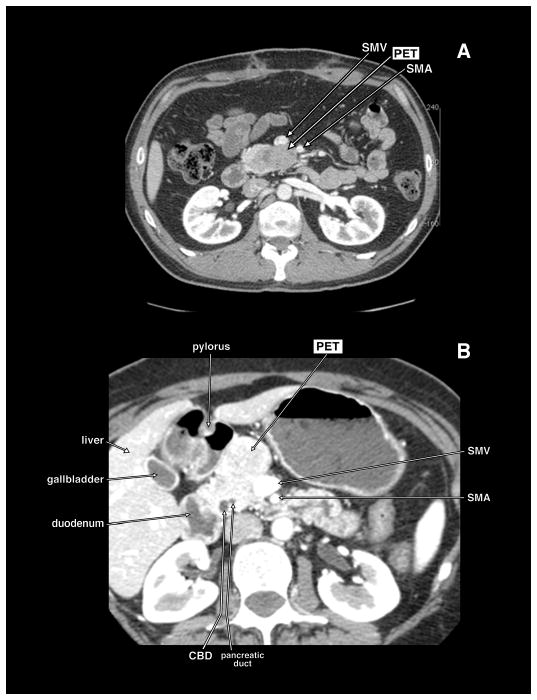
Figure 1.

Computed tomography (CT) scans of 2 different patients with pancreatic endocrine tumor (PET) in the head of the pancreas abutting the mesenteric vessels. In panel A, the PET is in the uncinate portion of the pancreatic head and lies abutting the posterior surface of the superior mesenteric vein (SMV) and superior mesenteric artery (SMA). In panel B, the PET is in the anterior portion of the head of the pancreas abutting the anterior and lateral wall of the SMV. These patients could have the PET dissected off the SMV.
