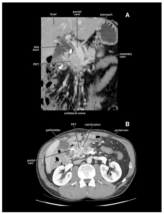
Figure 2.

Coronal planar reformation (panel A) and axial tomogram (panel B) of a computed tomography of the same patient with a locally invasive non-metastatic pancreatic endocrine tumor (PET) obstructing the proximal portal vein. The PET has calcifications. There are extensive collateral veins because of the portal vein obstruction. This patient had the portal vein resected and reconstructed with autologous femoral vein.
