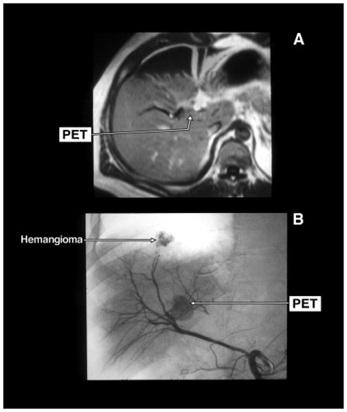
Figure 3.

Gadolinium enhanced magnetic resonance imaging (MRI) (panel A) and selective arteriogram (panel B) of a pancreatic endocrine tumor (PET) that was in the wall of the right hepatic duct. The tumor was abutting the right portal vein. There is a second liver tumor shown on the hepatic arteriogram (panel B) as a liver hemangioma. The PET was locally resected with the right hepatic duct. The tumor was dissected off the portal vein.
