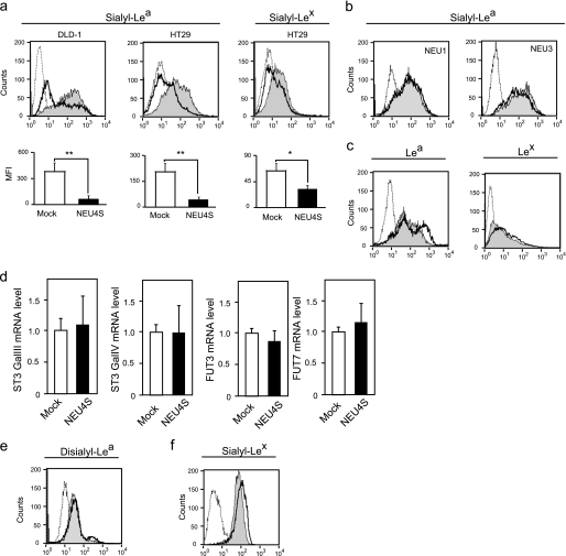
FIGURE 1.
Down-regulation of sialyl-Lea and sialyl-Lex in DLD-1 and HT29 colon cancer cells by NEU4S overexpression. a, flow cytometric analysis of cell surface sialyl-Lea in DLD-1 (left), HT29 (middle) cells, and sialyl-Lex in HT29 (right) (dotted line, mouse IgG; line filled with gray, Mock; black line, NEU4S). The data are mean ± S.D. from three experiments (*, p < 0.05; **, p < 0.01). b, effects of NEU1 (left) and NEU3 (right) overexpression on cell surface sialyl-Lea in HT29 cells (dotted line, mouse IgG; line filled with gray, Mock; black line, NEU1 or NEU3). c, altered expression of Lea (left panel in DLD-1) and Lex (right panel in HT29) by NEU4S (dotted line, mouse IgG; line filled gray, Mock; black line, NEU4S). d, relative expression of ST3 GalIII, ST3 Gal IV, FUT3, and FUT7 in NEU4-overexpressing HT29 cells. mRNA levels were assessed by real time PCR. e, no significant change in disialyl-Lea expression by NEU4S (dotted line, mouse IgG; line filled gray, Mock; black line, NEU4S). f, up-regulation of sialyl-Lex in HepG2 cells by NEU4S knockdown (dotted line, mouse IgG; line filled gray, scramble control; black line, NEU4S RNAi).

