FIGURE 7.
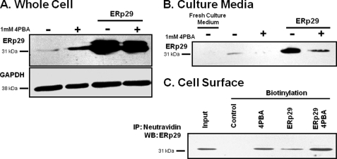
Detection of ERp29 in growth medium and at surface of IB3-1 cells. IB3-1 cells were grown under control conditions (−), in the presence of 1 mm 4PBA (+), transfected with 2 μg of ERp29, or transfected with ERp29 in the presence of 1 mm 4PBA for 24 h as indicated. A, whole-cell lysates were prepared, and ERp29 and GAPDH (as a loading control) were detected by immunoblot. Data are representative of n = 4 independent experiments. B, growth medium was collected and concentrated 10-fold as described under “Experimental Procedures.” ERp29 was detected in the conditioned medium by immunoblot but not in a similarly concentrated fresh culture medium control. Smaller or larger immunoreactive species were not detected. Immunoblots representative of n = 4 independent experiments are shown. C, surface proteins were biotinylated, captured with neutravidin beads, and resolved by SDS-PAGE as described under “Experimental Procedures.” ERp29 was detected in the biotinylated fraction. The “Input” lane represents the immunoreactivity of 10% of the total protein subjected to neutravidin capture in the Control lane. GAPDH immunoreactivity was absent from the biotinylated fraction (not shown), suggesting that intracellular proteins were not labeled by biotin in this experiment. These data are representative of n = 4 independent experiments. IP, immunoprecipitation; WB, Western blot.

