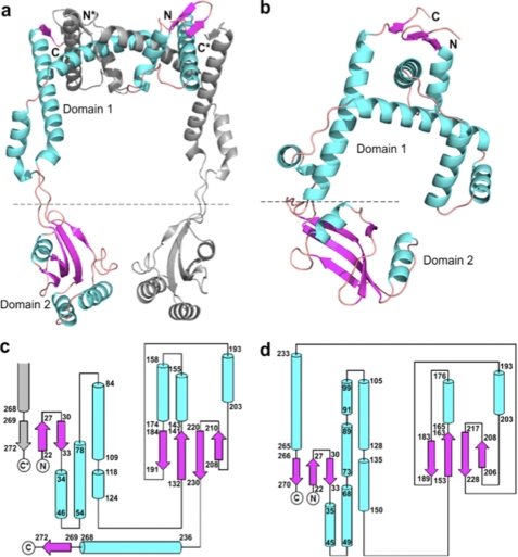
FIGURE 1.
The structures of PEB4 and Cj1289. a, a dimer of PEB4 as observed in the asymmetric unit of the crystal depicted in ribbon form with α-helices and β-strands shown as coils and arrows, respectively. One monomer is colored gray and the second monomer with helices in cyan and strands in magenta. The domains are indicated (domain 1 is the chaperone domain and domain 2 is the PPIase) and termini from the monomers are differentiated with *. Formation of an inter-monomer 3-stranded β-sheet can be clearly seen in the top right-hand corner and emphasizes the domain swapped architecture. b, a monomer of Cj1289 depicted in the same manner and with the same color scheme as in a. The equivalent 3-stranded β-sheet is clearly intra-monomer. c and d, topology diagrams of PEB4 and Cj1289, respectively. The α-helices and β-strands are shown as cylinders and arrows and the color scheme is maintained from a. Residue numbers marking the extent of the secondary structure elements are given. The PPIase domain 2 is shown on the right of each diagram. In c the formation of the inter-monomer β-sheet is indicated by the presence of an extra, gray colored strand from the other monomer in the dimer.

