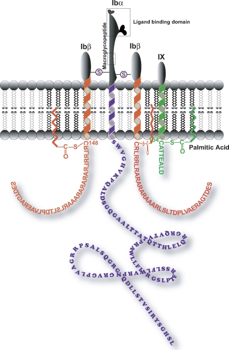
FIGURE 1.
Schematic view of the platelet GP Ib-IX complex. The depicted polypeptide arrangement of GP Ibα, GP Ibβ, and GP IX is based on the recently published stoichiometry of 1:2:1 (3). Circles with S inside represent the extracellular disulfide linkage between GP Ibα and GP Ibβ. The intracellular membrane-proximal cysteines of GP Ibβ (Cys148) and GP IX (Cys154) were reported to be the potential sites for palmitate modification as shown (15). The dashed rectangle depicts either the GP Ibα ligand binding domain or the WM23 binding region (macroglycopeptide).

