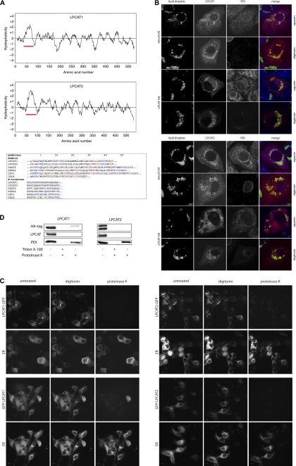
FIGURE 3.
LPCAT1 and LPCAT2 contain a long hydrophobic stretch and insert into LDs and membranes in a monotopic conformation. A, upper panels, hydrophobicity profiles of human LPCAT1 and LPCAT2 were calculated using the Kyte-Doolittle algorithm with a window of 17 amino acids. Red bar, N-terminal long hydrophobic stretch. Lower panel, sequence comparison between the hydrophobic sequences of LPCAT1 and LPCAT2 and the hairpin hydrophobic domains of the LD proteins DGAT2, NSDHL, and caveolin 1 (CAV1) and transmembrane domain of the ER protein calnexin (CALX). Position 1 indicates the first residue of the hydrophobic stretch or the N-terminal methionine. Color code in the membrane domains is as follows: charged residues, blue; helix-braking (Pro, Gly, Ser, and Cys), red. Color code for N termini: charged residues, blue; leucine, green. Sequences are taken from SwissProt with the following accession numbers: LPCAT1, Q8NF37; LPCAT2, Q7L5N7; DGAT2, Q96PD7; NSDHL, Q15738; CAV1, Q03135; CALX, P27842. B, A431 cells were transfected with vectors coding for LPCAT1 or LPCAT2 as indicated, both with either the N-terminal HA3 tag (HA-LPCAT) or C-terminal HA3 tag (LPCAT-HA) and cultivated in the presence of 100 μm oleate. Cells were fixed and processed for immunofluorescence microscopy using either saponin (permeabilizes all membranes) or digitonin (permeabilizes plasma membrane but not the ER membrane) for permeabilization. Cells were stained with Bodipy 493/503 to stain LDs (green), anti-HA to detect transfected LPCAT proteins (red), and anti-PDI (blue) as a lumenal ER marker (scale bar, 10 μm). C, living A431 cells overexpressing LPCAT1-GFP or GFP-LPCAT1 (upper panel) or LPCAT2-GFP or GFP-LPCAT2 (lower panel), all cultivated in the presence of oleate, were treated with digitonin. Cells were exposed to proteinase K, and images were recorded. Representative images before treatment (untreated), after 1 min digitonin (digitonin), and after 30 s of proteinase K exposure (proteinase K) are shown. The integrity of the ER at all conditions was monitored via the lumenal DsRed2-ER (ER). Scale bar, 10 μm. D, A431 cells expressing N-terminally HA3-tagged LPCAT1 or LPCAT2 were lysed, and microsomes were isolated. Microsomes were incubated in PBS with either no addition (negative digestion control), addition of 1% Triton X-100 and proteinase K (positive digestion control), or addition of proteinase K alone. The integrity of the microsomes was analyzed by Western blotting against the lumenal ER marker PDI. The N terminus of LPCAT1 and LPCAT2 was detected with a HA-specific antibody and the C terminus with LPCAT1 and LPCAT2 antibodies. No smaller fragments were detected on the blots (data not shown).

