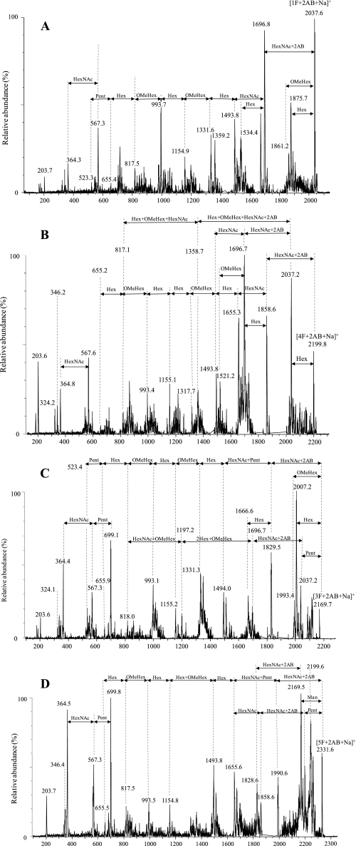
FIGURE 5.
Positive ion MS/MS spectra of the 2AB-labeled N-glycans released from the 66-kDa glycoprotein by PNGase F. Shown are glycan at m/z 2038 ([1F+2AB+Na]+ ion) (A), at m/z 2200 ([4F+2AB+Na]+ ion) (B), positive ion MS/MS spectra of the glycan at m/z 2170 ([3F+2AB+Na]+ ion) (C), and at m/z 2332 ([5F+2AB+Na]+ ion) (D). The arrows on the spectra indicate mass differences corresponding to glycan residues and do not necessarily imply a fragmentation sequence.

