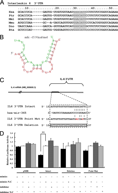
FIGURE 2.
Identification of the binding site for miR-365 in the IL-6 3′-UTR. A, alignment of the IL-6 3′-UTR showed that the MRE (gray) was conserved among different species. B, shown is a schematic representation of the hybridization between miR-365 and IL-6 3′-UTR. Green and red letters indicate miR-365 and IL-6 mRNA, respectively. C, shown is a schematic representation of mutant reporters of IL-6 3′-UTR. The frame and red letters indicate the deletion and the point mutation, respectively. D, intact, point mutant, or deletion mutants pMIR-IL-6 3′-UTR and pMIR-REPORT were co-transfected with the indicated oligonucleotides into HEK293 cells. 24 h post-transfection, luciferase activities were measured. Results are presented as -fold induction over control groups. Bar graph data are presented as the means ± S.D. (n = 3); **, p < 0.01, as compared with the transfection with the scrambled oligonucleotides.

