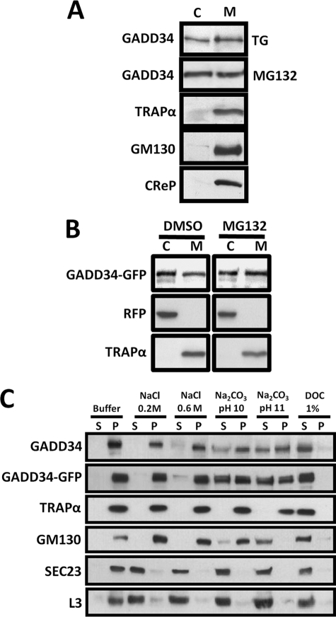
FIGURE 1.
Subcellular distribution of human GADD34. A, HeLa cells subjected to ER stress by exposure to 1 μm thapsigargin (TG) or 5 μm MG132 for 5 h were homogenized as described under “Experimental Procedures.” Parallel samples of 100,000 × g supernatant or cytosol (C) and membrane pellet (M) were subjected to SDS-PAGE and immunoblotting with anti-GADD34, anti-TRAPα, anti-GM130, and anti-CReP antibodies. B, HeLa cells coexpressing GADD34-GFP and RFP were treated with either DMSO or 5 μm MG132 for 5 h and subjected to fractionation as described in panel A, and the cytosol and membrane fractions were immunoblotted with anti-GFP, anti-RFP, and anti-TRAPα antibodies. C, purified membranes from HeLa cells exposed to 1 μm thapsigargin for 5 h to induce endogenous GADD34 or expressing WT GADD34-GFP were incubated in a variety of extraction buffers as described under “Experimental Procedures.” Following centrifugation, supernatant (S) and pellet (P) were subjected to SDS-PAGE and immunoblotting with anti-GADD34, anti-GFP, anti-TRAPα, anti-GM130, anti-SEC23, and anti-ribosomal L3.

