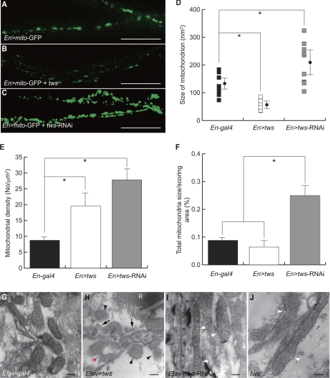
FIGURE 3.
Tws affects the morphogenesis of mitochondria in CNS. A, mitochondria in the axons of ventral ganglia were labeled with mito-GFP driven by En-gal4. B, fragmentation of mitochondria in En>tws flies. C, fusion of mitochondria in En>tws-RNAi flies. Scale bar, 20 μm. D–F, Tws regulates the size, density, and biogenesis of mitochondria. Statistical analyses were performed using one-way analysis of variance with supplementary Student-Newman-Kewls test and are expressed as mean ± S.D.; * indicates p < 0.001. G–I, ultrastructure of mitochondria in the transverse sections of adult retina was accessed by transmission electron microscopy. Normal mitochondria in Elav-gal4. H, ectopic Tws caused scission of mitochondria (red arrowhead). Many disintegrated mitochondria with less condensed cristae were present (black arrowheads), and some were circled by autophagosome-like organelles (arrows). I and J, tubular mitochondria with swollen cristae were present in Elav>tws-RNAi or tws mutant (twsp flies (white arrowheads). R = rhabdomere. Scale bar, 200 nm.

