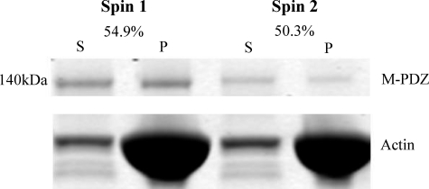
FIGURE 7.
Evidence for two conformational states of myosin-18 motor. Myosin-18-MMD proteins were sedimented with 20 μm actin as in Fig. 6. The left two lanes show the supernatant (S) and pellet (P) fractions from a representative experiment using M-PDZ. The supernatant from this experiment was mixed with 20 μm actin, and a second sedimentation was performed. The supernatant and pellet from this experiment are shown in the right two lanes. The fraction of actin bound in the first sedimentation was 54.9%, and the fraction bound in the second sedimentation was 50.3%. Ionic conditions were as described in Fig. 6.

