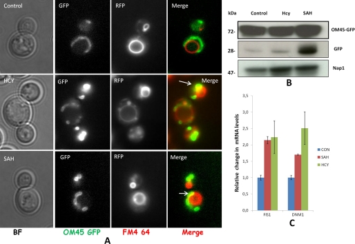
FIGURE 9.
Monitoring mitophagy response in str4Δ strain. A, co-localization of mtGFP and vacuoles. str4Δ strain containing OM45 protein tagged with GFP as the C-terminal tag was grown in the presence and absence of 5 mm homocysteine and 600 μm AdoHcy for 12 h. To stain the vacuole, FM4-64 (at a final concentration of 40 μm) was added for 2 h. Live cells were examined under a fluorescence microscope in their respective media to check proper vacuolar staining and Om45-GFP in live cells. Images were acquired with epifluorescence microscope and analyze by ImageJ software. B, detection of free GFP by immune blotting. Cells were collected at 12 h, lysed, and subjected to Western blot analysis with anti-GFP antibody or Nap1p antibody (loading control). The positions of full-length Om45-GFP and free GFP are indicated. C, mitochondrial fission gene expression. Cells were grown in the presence and absence of homocysteine and AdoHcy (SAH) for 16 h. RNA was isolated from these cells and subjected to real time PCR analysis using the gene-specific primers. IPP1 (inorganic pyrophosphatase) gene was used as the internal control (CON) gene for normalization. Error bars are representative of means ± S.D. (n = 2).

