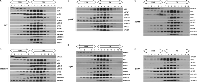
FIGURE 2.
Membrane sublocalization of different PSII assembly factors. Synechocystis cell extracts of the wild-type strain (A) and pratA− (B), ycf48− (C), ins0933 (D), ctpA− (E), and psbA− (F) mutant strains were separated by two consecutive rounds of sucrose density gradient centrifugation (5). The second linear gradient from 20 to 60% sucrose was apportioned into 14 fractions, which were analyzed by immunoblotting using the indicated antibodies. Fractions 1–6 represent PDMs, and fractions 7–14 represent TMs. To facilitate comparison between gradients, sample volumes were normalized to the volume of fraction 7 that contained 40 μg of protein. Due to the sharp fall in the levels of pD1-containing RC complexes in ins0933, no pD1 signal could be detected in this mutant (15). Because no mature D1 can accumulate in the ctpA− mutant, pD1 was detected using anti-D1 antiserum. For the pD1 signal of pratA−, see Ref. 5.

