
Figure 1.
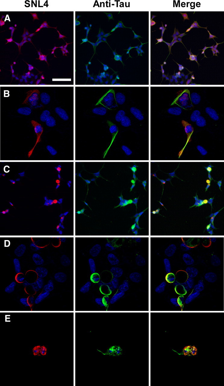
Tau colocalized with α-syn in cells that form cellular aggregates. Representative double immunofluorescence between SNL4 (anti-α-syn antibody) and an anti-tau antibody was performed on QBI293 cells cotransfected with expression plasmids for human wild-type α-syn and tau. A, B, In the absence of fibril treatment, α-syn appeared diffuse, and tau appeared diffuse and bundled. C–E, For cultures treated with recombinant, prefibrillized 21–140 α-syn protein, intracellular, endogenously generated α-syn formed large cellular aggregates that sometimes contained tau. Colocalization of α-syn and tau was assessed by confocal microscopy (B, D, E). Overlay between a portion of SNL4 and anti-tau immunoreactivity was observed in aggregate-containing cells. Many of these cells appeared rounded in morphology, and occasional tau “tails” were observed attached to aggregated α-syn proteins. Representative images are of cells fixed 72 h after transfection. Equivalent exposures were provided for all representative samples. Scale bar: A (for A, C) 100 μm, (for B, D, E) 30 μm.