Figure 8.
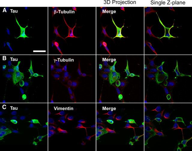
Confocal microscopy analyses of cytoskeletal markers on cells containing tau aggregates. A–C, QBI293 cells were transfected with expression plasmids for WT (A, C) or P301L (B) tau and treated with recombinant, prefibrillized full-length α-syn. Cells containing tau aggregates were identified by the characteristic increased anti-tau immunoreactivity and displacement of the nuclear membrane. Representative images are cells fixed 72 h after transfection. Double immunofluorescence was performed between anti-tau antibodies (green) and anti-β-tubulin (A), anti-γ-tubulin (B), or anti-vimentin (C) (red). Three-dimensional projections of cells were composed by stacking 10–12 confocal images and rotating images 15–30° on a central axis (first three columns). A single, merged Z-plane (<0.7 μm) of each representative image is provided at the far right column. Anti-β-tubulin (A) and anti-vimentin immunoreactivity (C) were observed around and inside some tau aggregates. Anti-γ-tubulin immunoreactivity (B) was observed outside tau aggregates, often displayed outside of the center of the cellular plane. Similar morphology of tau aggregates were observed for wild-type and P301L-expressing cells. Scale bar, 40 μm.

