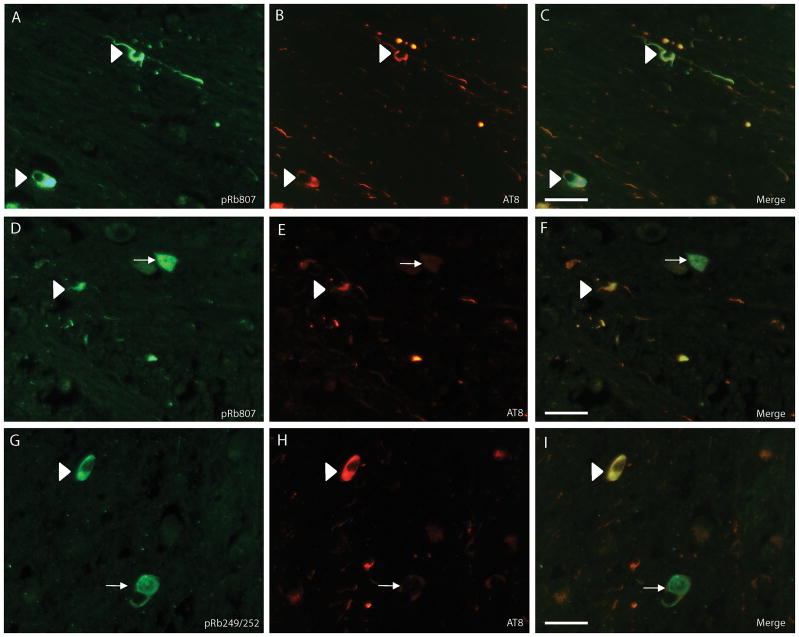
Figure 2.
Double label fluorescence microscopy in progressive supranuclear palsy (PSP). (A-C) A large number of PSP tangles show both ppRbser807 (A, green) and AT8 (B, red) marked with white arrowheads and there are also many AT8-positive neurites that do not contain ppRbser807 (C, merged image). (D-F) There are a few neurons that are strongly immunoreactive for ppRbser807 (D, green), but lack AT8 (E, red) (white arrow) and evident in merged image (F). White arrowheads indicate neurons with colocalization. (G-I) The same pattern is found using an antibody specific for ppRbS249/T252 (G, green). Many neurites only stain with antibody AT8 (H, red); some PSP tangles display only ppRbS249/T252 and not phosphorylated tau (I, merged image, white arrow); some PSP tangles show colocalization (white arrowheads). Scale bars = 50 μm.

