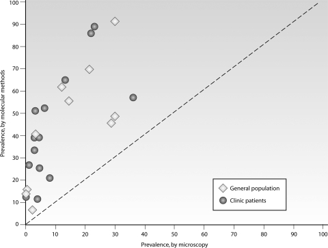
Fig. 4.
Gametocyte carriage by microscopy and molecular detection tools. Gametocytes were detected by microscopy, typically screening 100 microscopic fields, and by pfs25- or pfg377-specific RT-PCR, LAMP, or QT-NASBA. Samples were derived from the general population in Burkina Faso, Tanzania, the Gambia, and Thailand (open diamonds) (70, 263, 319, 335, 337, 340, 413) and from people attending clinics in Kenya, Tanzania, Sudan, and Vietnam (closed circles) (1, 55, 140, 256, 279, 314, 412), mostly children participating in clinical trials. (Reproduced from reference 334 with permission.)

