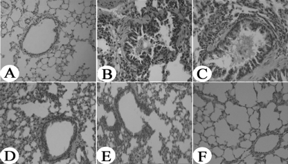
Fig. 4.
Reduced lung inflammation in high-dose OVA-CTLA-4-pcDNA3.1-vaccinated mice. Histological findings of lung tissues (original magnification, ×200) in mice from the control group (A), the model group (B), the pcDNA3.1 group (C), the OVA-pcDNA3.1 group (D), the OVA-CTLA-4-pcDNA3.1(L) group (E), and the OVA-CTLA-4-pcDNA3.1(H) group (F) were analyzed by using H&E staining.

