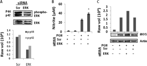
Fig. 5.
Knockdown of ERK1/2 augmented PGN-induced iNOS expression and NO production. Macrophages were transfected with ERK1 and ERK2 siRNA (ERK) or scrambled siRNA (Scr). After 36 h of transfection, macrophages were stimulated with PGN (10 μg/ml) for 30 min. (A) Phosphorylated ERK and total ERK protein were detected by immunoblotting with antibody specific for phospho-ERK (p-p42 and p-p44) or ERK2. The lower panel shows the densitometry analysis of phospho-ERK bands. (B) After 36 h of transfection, macrophages were stimulated with PGN (10 μg/ml) for 18 h, and the NO in the culture supernatant was evaluated by Griess reagent assay. Each bar represents the standard error of three independent experiments. *, P < 0.05 compared to scramble siRNA. (C) After 18 h, PGN-treated macrophages were lysed, and the cell lysates were immunoblotted with anti-iNOS polyclonal antibody. The same blot was reprobed with anti-actin antibody to demonstrate equal protein loading.

