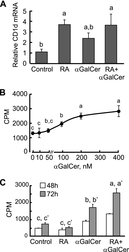
Fig. 1.
Regulation of CD1d expression and cell proliferation by RA and αGalCer in mouse splenic B cells. (A) RA increased CD1d expression in spleen B cells. B cells were cultured in the presence or absence of RA (20 nM) or αGalCer (100 nM) for 24 h. Total RNA was extracted and subjected to quantitative PCR analysis. The data are presented as the ratio of CD1d to tubulin mRNAs, representing three independent experiments with treatments in triplicate. a > b, P < 0.05. (B) αGalCer increases B cell proliferation dose dependently. B cells were isolated and cultured in the presence of different concentrations of αGalCer for 72 h. [3H]thymidine was added for the last 6 h of culture to monitor cell proliferation activity. a > b > c, P < 0.05. (C) B cells were isolated and cultured in the presence or absence of αGalCer and/or RA or anti-μ antibody (0.1 μg/ml). [3H]thymidine was added on day 2 or 3 for the last 6 h of culture to monitor cell proliferation activity. αGalCer strongly increased B cell proliferation, which was further enhanced by RA. a > b > c (48 h) and a′ > b′ > c′ (72 h), P < 0.05.

